Discovering a lump on your pet is understandably worrying, but finding it early is one of the most important things you can do. Whether the mass turns out to be a harmless fatty lump or something that needs treatment, the only way to know for certain is through diagnostic testing. The two main options are a fine needle aspirate (FNA, a straightforward procedure where a thin needle draws cells from the lump for examination under a microscope) and a biopsy (where a tissue sample is surgically removed and sent to a veterinary pathologist for a more detailed assessment called histopathology).
At The Vale Veterinary Group, we have in-house laboratory capabilities and diagnostic services across our Devon practices that allow us to investigate lumps and masses thoroughly and move toward answers quickly. Our team will talk you through the reasoning behind each recommendation so you understand what we are testing for and why. If you have noticed a new lump on your pet or a change in one you have been monitoring, contact us to arrange an appointment at your nearest branch.
Why Examination Alone Is Not Enough
Signs of cancer in pets do not follow a predictable appearance. Malignant tumours can feel soft and moveable; benign masses can feel firm and fixed. Size, texture, and growth rate all provide useful clinical clues, but none of them are diagnostic on their own.
Cancer in pets is common, particularly as animals age. Earlier diagnosis consistently produces more treatment options and better outcomes. The decision to test a lump promptly is almost always worthwhile.
Fine Needle Aspirate: The First Diagnostic Step
What the Procedure Involves
A fine needle aspirate uses a thin needle to collect cells from within the mass. These cells are spread onto a slide and examined under a microscope, a process called cytology. The procedure typically takes a few minutes, does not require sedation for most pets, and feels similar to a vaccination. Multiple sites can be sampled in the same appointment if more than one mass is present, and results from our reference laboratory usually return within a few working days.
What FNA Can Identify
Cytology is most informative for well-defined tumour types and conditions:
- Lipomas: fat-containing cells confirm the benign fatty origin
- Mast cell tumours: characteristic granules visible under the microscope are strongly diagnostic
- Cysts: fluid content and cell type confirm the nature of the lesion
- Reactive versus enlarged lymph nodes: cytology often distinguishes these directly
When FNA Falls Short
Some tumour types release cells poorly into a needle, producing non-diagnostic samples even when a significant mass is present. Fibrosarcomas, some mammary tumours, and certain connective tissue tumours are examples. A growing or suspicious mass with a non-diagnostic FNA result should proceed to biopsy rather than further monitoring.
Skin Masses That Look Similar but Behave Very Differently
One of the strongest arguments for testing rather than guessing is how often masses with completely different prognoses look nearly identical from the outside. A small pink lump might be a self-resolving condition or an aggressive cancer; the appearance alone does not say which.
Commonly Confused Skin Masses in Dogs
The masses most likely to cause confusion in dogs include:
- Histiocytomas: small, raised, often hairless red lumps that appear suddenly in young dogs. Almost always benign and frequently resolve on their own within a few months. Can look identical to a mast cell tumour on inspection.
- Melanocytic tumours: pigmented masses ranging from benign moles to aggressive melanomas, particularly concerning when found in the mouth or near nail beds.
- Cysts in dogs: fluid- or sebum-filled lumps that feel firm and well-defined, often mistaken for tumours until aspirated.
- Mast cell tumours: notoriously variable in appearance, sometimes mimicking insect bites, lipomas, or histiocytomas. Grade and behaviour cannot be predicted by appearance.
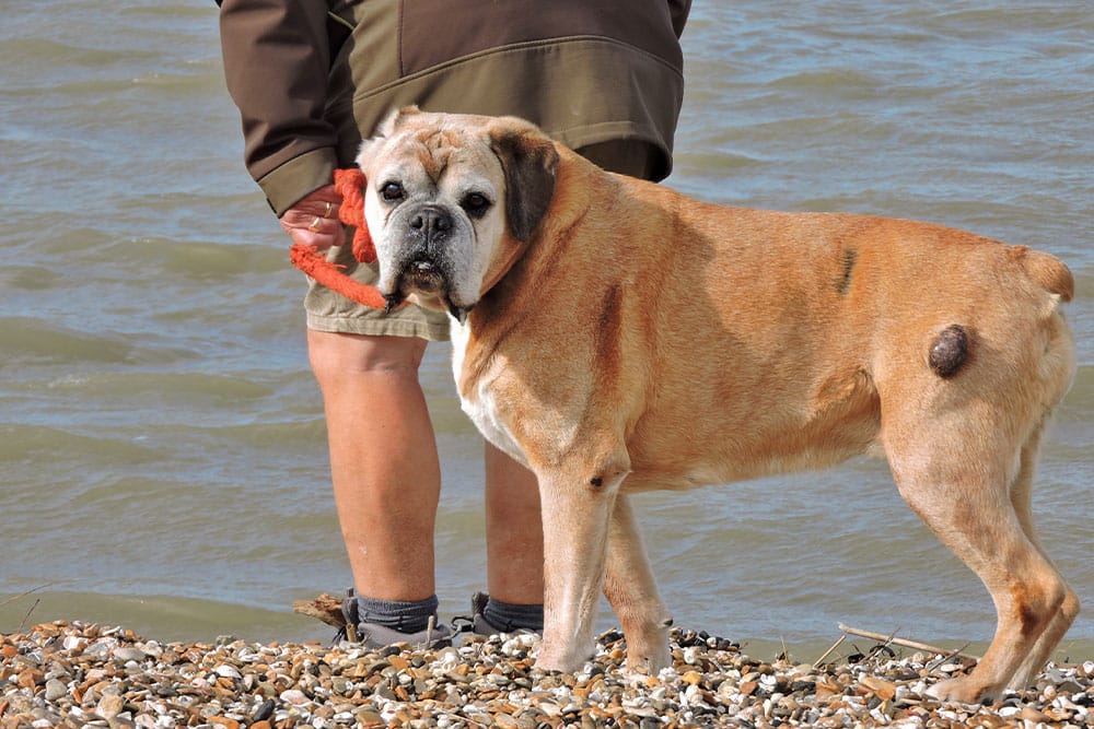
- Lipomas: soft, moveable, slow-growing fatty masses, common in middle-aged and older dogs, but worth confirming because the more aggressive infiltrative lipoma and the malignant liposarcoma can feel similar. Lipomas often grow so large that they impede movement, so even if benign, removal may be warranted.
The visible features simply do not separate these reliably enough to skip testing.
Commonly Confused Skin Masses in Cats
In cats, the stakes of misidentification tend to be higher because cats carry a greater proportion of malignant masses than dogs do.
- Feline basal cell carcinoma: typically firm, raised lumps on the head, neck, or shoulders. Often slow-growing and locally invasive but rarely spreading to distant sites.
- Squamous cell carcinoma: crusty, ulcerated, or non-healing lesions, particularly on the ears, nose, and eyelids of white or light-coated cats with sun exposure. Aggressive when not caught early.
- Fibrosarcoma in cats: firm subcutaneous masses, sometimes at injection sites, that infiltrate surrounding tissue extensively. Wide surgical margins are needed.
- Feline mast cell tumours: present as either solitary skin lumps (often benign) or visceral disease affecting the spleen and intestines (more serious). The skin form is far more common in older cats.
For any new lump in a cat, prompt cytology is the standard recommendation rather than watchful waiting.
Biopsy and Histopathology
How Biopsy Works
A biopsy removes a tissue sample rather than individual cells, allowing the pathologist to evaluate cellular organisation, invasion patterns, tissue architecture, and margin status. These features are only visible in sections of intact tissue and cannot be assessed through cytology alone.
Common biopsy approaches:
- Punch biopsy: a circular cutting instrument removes a cylinder of tissue; suitable for well-defined superficial masses
- Incisional biopsy: a wedge of tissue is removed without excising the whole mass; used when diagnosis before definitive surgery is needed
- Excisional biopsy: the entire mass is removed and submitted, combining treatment and diagnosis
- Core needle biopsy: a larger-gauge needle collects a tissue core preserving architectural detail
All biopsy procedures are performed under sedation or general anaesthesia. Tumour diagnosis through histopathology provides what cytology alone cannot.
What Histopathology Establishes
The veterinary pathologist evaluates the biopsy to determine:
- Tumour type: definitive identification of the specific cancer or benign condition
- Grade: how aggressive the cells appear histologically
- Invasion: whether the tumour is infiltrating surrounding tissue
- Margins: whether surgically excised tissue has clear edges or tumour present at the cut margin
For the types of cancer that look identical on cytology but behave entirely differently, histopathology resolves the ambiguity that guides treatment decisions.
When Biopsy Is the Only Way to Get a Reliable Answer
Some clinical scenarios cannot be resolved by cytology alone, and proceeding to biopsy is the right call from the outset.
Splenic Masses: Benign or Haemangiosarcoma?
A mass on the spleen, found incidentally on imaging or during the workup of a collapsed pet, presents one of the most challenging diagnostic situations in small animal medicine. Imaging cannot reliably distinguish a benign splenic haematoma from haemangiosarcoma, an aggressive malignant tumour of blood vessel origin. FNA of splenic masses is often unreliable because the samples can contain mostly blood rather than diagnostic cells, and the cells that are present do not always reveal the underlying disease.
In most cases, the definitive answer comes from removing the spleen (splenectomy) and submitting the tissue for histopathology. This produces a clear diagnosis and, when the mass is benign, may also be the complete treatment.
Chronic GI Disease in Cats: IBD or Lymphoma?
Inflammatory bowel disease in cats and intestinal lymphoma can produce nearly identical clinical signs: chronic vomiting, weight loss, soft stools, and reduced appetite. Bloodwork and imaging often look similar between the two conditions, particularly with the low-grade form of intestinal lymphoma.
The treatments, however, are entirely different. IBD is managed with diet changes and immunosuppressive medication; lymphoma requires chemotherapy. Endoscopic or surgical biopsy of the intestinal wall is the only reliable way to differentiate them, which is why we recommend tissue sampling rather than empirical treatment for cats with persistent GI signs.
Liquid Biopsy and Blood-Based Cancer Screening
Newer blood-based cancer screening tests detect cancer-associated DNA fragments in the bloodstream, sometimes before a visible mass develops. They are most useful as a screening tool for older pets and breeds with documented cancer predispositions, including lymphoma in Golden Retrievers and other high-risk lines.
A positive screening result is not a diagnosis on its own. It signals that further workup is appropriate: imaging, additional bloodwork, and targeted aspirates or biopsies based on what is found. A negative result reduces the immediate concern but does not rule out cancer entirely. Liquid biopsy fits naturally alongside routine healthcare for senior pets and is something we can discuss as part of annual screening for high-risk patients.
Choosing Between FNA and Biopsy
FNA is typically the first step because it is fast, low-cost, and non-invasive. Biopsy is recommended when:
- FNA returns non-diagnostic from a suspicious mass
- Tumour grading is needed to guide treatment planning
- Margin assessment after excision is clinically important
- Clinical suspicion for malignancy remains despite a benign FNA result
Our team discusses these trade-offs openly at the appointment so you understand the basis for the recommendation and what each result will and will not tell us.
Monitoring Benign Masses Over Time
A benign cytology result does not end the conversation. Masses can change behaviour, new masses can appear, and the original assessment was a snapshot of one moment in time. A simple monitoring routine catches changes early:
- Measure the mass with a soft tape or callipers and record the dimensions
- Photograph it next to a coin or ruler for scale, in consistent lighting
- Check monthly with a quick visual review and a gentle palpation
- Note any change in size, texture, surface, or whether it has become bothersome to your pet
Reasons to call us between scheduled visits include noticeable growth over four to eight weeks, change in texture or consistency, any discharge, ulceration, or surface change, a new mass appearing nearby, or any mass on a cat regardless of how it looks.
What Happens After Results Come Back
Results generally fall into one of three categories, each of which guides what comes next:
- Benign: in most cases this means a monitoring plan, with the option of removal if the mass becomes a nuisance, grows quickly, or sits in a location where it bothers your pet.
- Malignant: opens a treatment conversation that may include surgical excision with appropriate margins, referral for further imaging or oncology consultation, and discussion of grade-specific options.
- Inconclusive: points us toward the next diagnostic step, which is usually biopsy, repeat aspiration, or imaging to better characterise what we are dealing with.
In every case, our team explains what the results mean in plain language and outlines next steps clearly so you are not left to interpret a laboratory report on your own. A great resource is the Animal Cancer Trust\- they have information on cancer types and case studies to help better understand what’s happening with your pet.
What the Process Looks Like at The Vale
For FNA: a brief appointment at any of our Devon branches. The area is gently prepared, the sample is collected over a minute or two, and your pet returns home the same day. Results typically return within a few working days.
For biopsy: pre-anaesthetic assessment and any indicated bloodwork. The procedure under sedation or general anaesthesia. Discharge with an e-collar and appropriate pain management. Suture removal at ten to fourteen days. Histopathology results within one to two weeks.
Our small animal team explains findings in clear language and outlines next steps directly so you are not left to interpret a laboratory report without support.
Frequently Asked Questions
Can I just keep an eye on the lump and see what happens?
For some low-risk masses, monitoring with documented size checks is appropriate. For any mass that is growing, changing, or causing clinical signs, testing provides information that monitoring cannot.
My pet had an FNA and the result was benign. Is that conclusive?
For many well-defined mass types, yes. For masses where clinical suspicion remains despite a benign result, or where the FNA returned non-diagnostic, biopsy provides more definitive information.
How long do results take?
FNA cytology results typically return within two to five working days from our reference laboratory. Histopathology from biopsy samples usually takes one to two weeks.
Does my pet need to be sedated for an FNA?
Most pets tolerate FNA without sedation, particularly when the mass is in an accessible location. For anxious pets, lumps near sensitive areas, or deeper masses requiring imaging guidance, light sedation is sometimes appropriate and we discuss this case by case.
Getting to a Clear Answer
At The Vale Veterinary Group, our in-house diagnostic capabilities and our experienced clinical team mean we can move from an unexpected lump to a clear plan quickly. The right test, performed at the right time, is what turns uncertainty into a path forward.
Contact us to arrange an appointment at your nearest branch.


Leave A Comment